
At Shoal Creek Foot & Ankle Center, we use high-frequency digital X-ray technology to deliver fast, precise imaging with minimal radiation exposure. Our advanced system produces crystal-clear images while allowing for quick, comfortable patient positioning — so we can diagnose issues accurately and keep your visit efficient.
What to Expect During an X-Ray
Getting an X-ray is a quick and painless part of your visit. Here’s what usually happens:
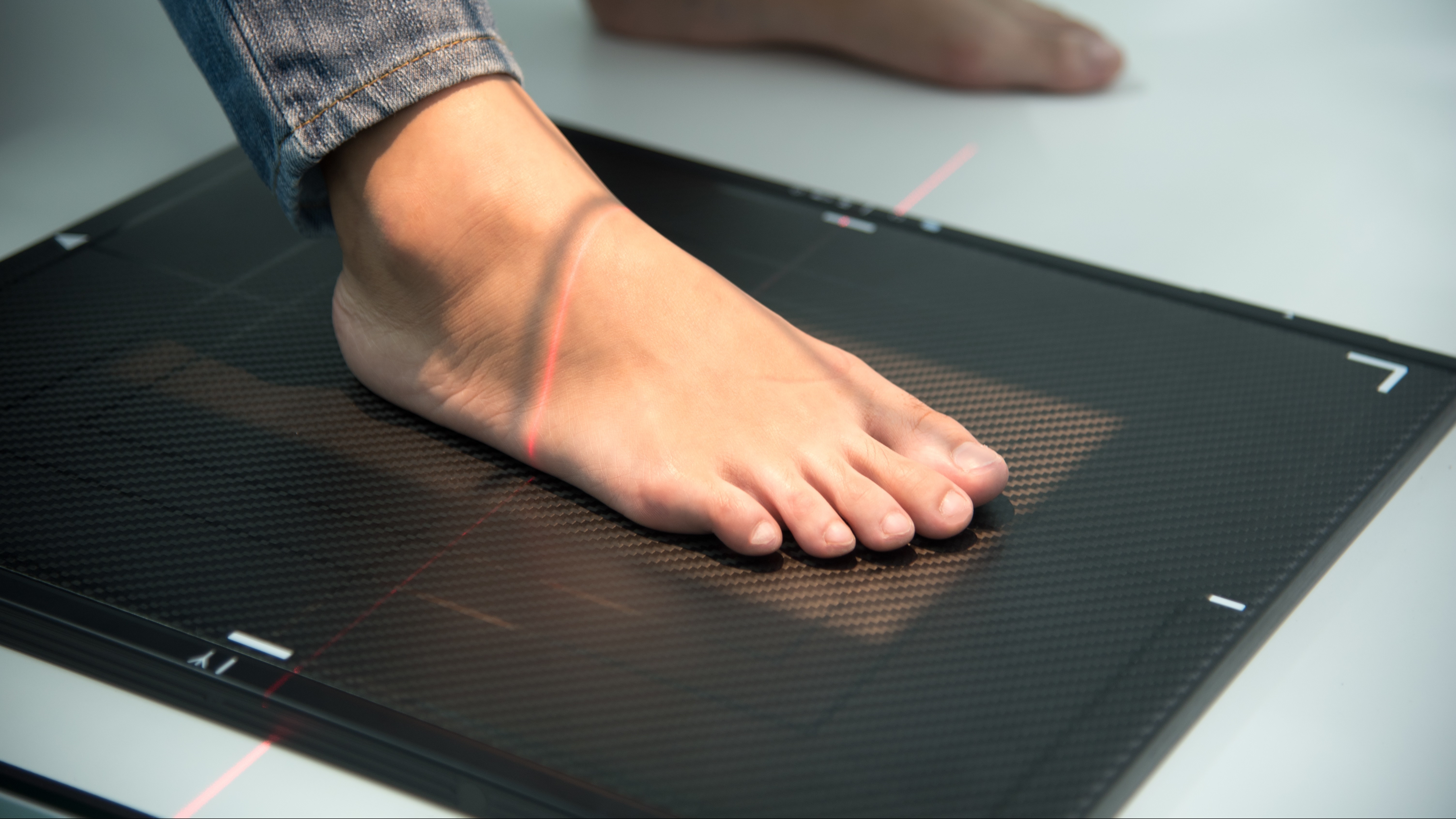
- One of our Medical Assistants will help position your foot or ankle on the X-ray platform.
- Your foot will be aligned between the X-ray machine and detector.
- You’ll need to hold still for a few seconds while the image is taken.
- We may take several views to get the most accurate look at what’s going on.
Once the images are complete, your podiatrist will review them with you in the exam room and explain the findings.
Why Weightbearing X-Rays Matter
Weightbearing (standing) X-rays are a game-changer when it comes to foot and ankle care. They show how your bones and joints align while you’re actually standing and bearing weight — which gives us a much more accurate picture than lying-down images ever could.
These images help diagnose:
- Fractures and dislocations
- Midfoot and forefoot injuries
- Bunions
- Flat feet and fallen arches
- Sprains, strains, and instability
- Arthritis and joint degeneration
- Diabetic foot changes
They’re also critical for surgical planning — helping our surgeons place screws, plates, or implants exactly where they need to go, which leads to better outcomes and faster recovery.
